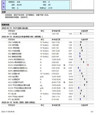
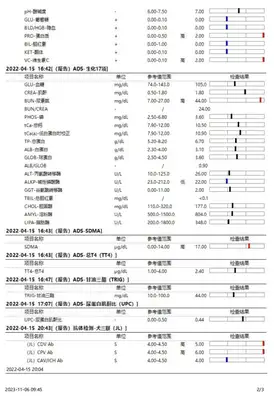
2. Basic information 2.2. Disease history & hospital examination results 2.2.2. Results of examination results in March 2023 2.2.3. Examination results in April 2023 2.3.
Dog testicular tumours are a disease occurring in dogs that is more hairy than in other animals, especially in certain varieties, such as fighting lions, Chihuahuas, Pomenanias and precious dogs, but most of them are found in geriatrics.
Clinical manifestations of testicular neoplasms are mainly the swelling of the testicles, which may grow larger and hard, but usually do not cause pain. As tumours may be administered with estrogens, feminized behaviour may occur in dogs, such as constriction, attraction to other dogs, etc.
The canine testosterone tumours are mainly classified as sexual mesothelioma (Sex Cord Stromal Tumors) and reproductive cell tumours (Germ Cell Tumors), with a further breakdown in support of Sertoli Cell Tumors, Interstitial Cell Tumors and Seminoma.
This case was reported as an inter-cell tumor in the testicle. Basic information on the content of cases
Pippi:
Siberian sleigh dogs.
8 years (approx. 50 years)
Male, unsterilized
22.60 kg
Situation rating, Bcs (Body Consultation Scring): 4/9, thin
CRT (Creaconary refilling time): 1-2s
CKD (chronic kidney disease): II
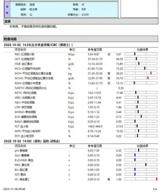
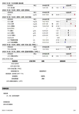
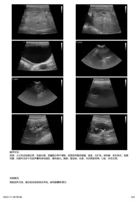
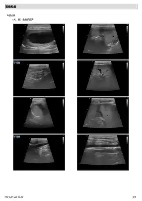
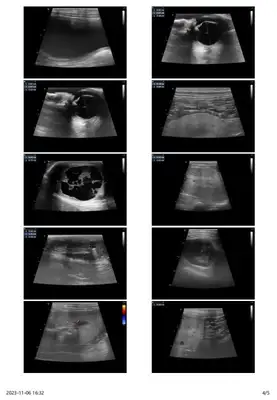
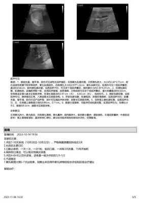
The principal has recently found that the two testicles vary considerably in size, mental state and selection. The results of the ultrasound examination of the pathological parts of the hospital were as follows:
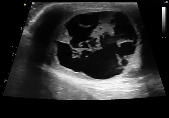 This is a swollen left testicle.
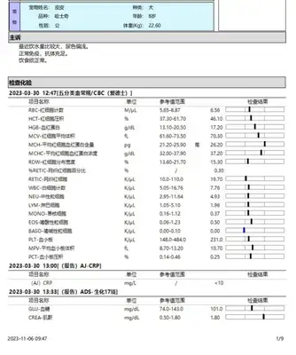
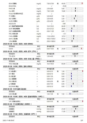
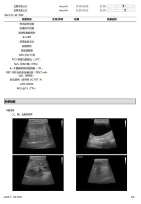
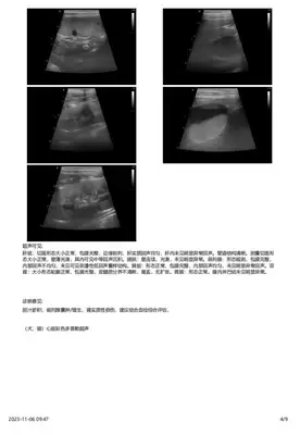
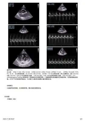
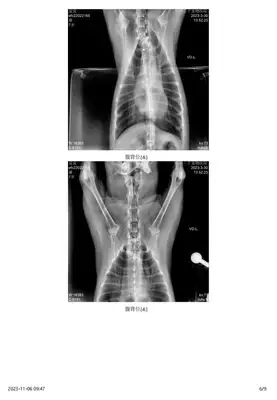
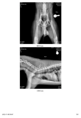
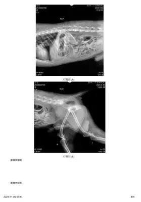
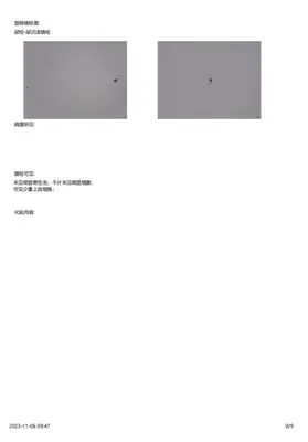 Results from April 2023
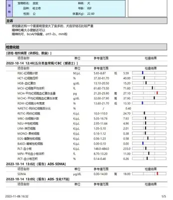
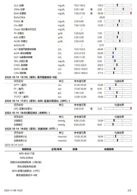
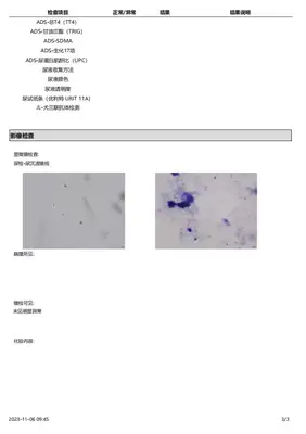 October 2023 Inspection results
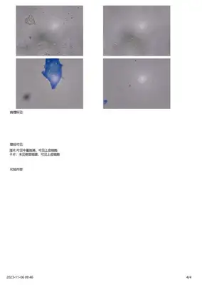
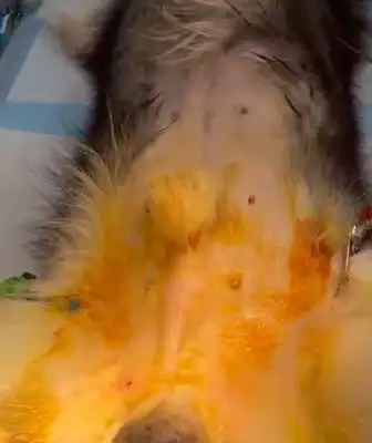
 Surgery photos
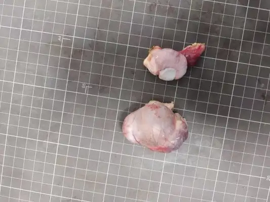
The latter two testosterone tissues were given to the M.E.L. for the organization of pathological examinations using the Formalin fixed. General pathological diagnosis and collection of photographs
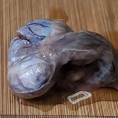
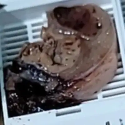
The screening organization is a testicle and an affiliate. Eyes are grey red, with a size of 6.5* 5.4*4.2 cm; 5.2* 3.8*3.5 cm. In tissue, the profile is red and red. A cross-section with a total of 4 pieces of tissue, in 4 groups.
 Mirror Description
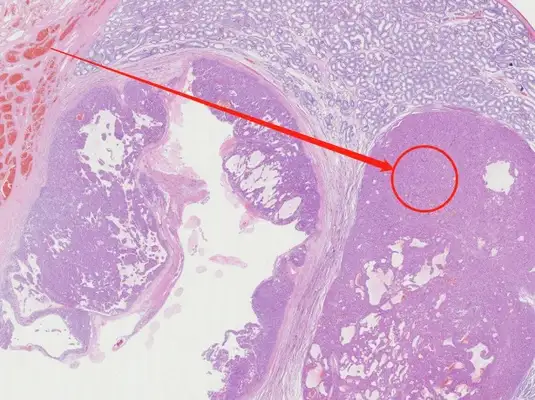
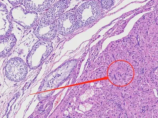
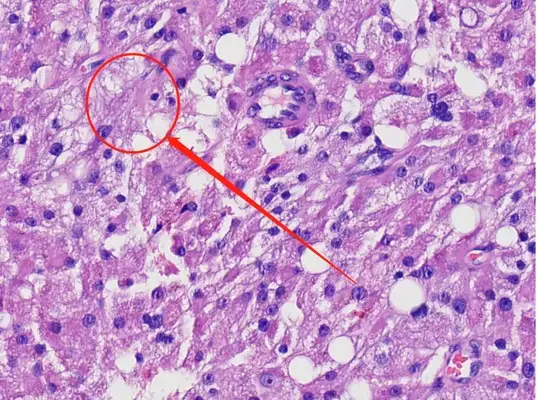
The testosterone sample is 2 pieces, visible under the mirror: both testosterone have the same pathological manifestations. Tumor cells are located in the centre of the swelling and are found in tablets and irregular glands. These cells have a smaller nucleus of deep infection, occasional nucleotide, and a smaller heterogeneity of cell nucleus. Cells are acidic and in large quantities, and some of the cells have a significant concentration of lipid. The whole cell is of a small heterogeneity, with no filamental cleavage in an average of 10 hyperscopes. Cream vessels are highly active, with severe blood silt in some areas.
 Pathological diagnosis
Leydig Cell Tumor, completely removed.
Abundance cut: Yellow brown, soft, cut up. They usually contain bleeding and/or cysts. These are consistent with the results of clinical ultrasound skepticism.
For inter-cellular tumors in dogs' testicles, decomposition is curable. Discussion
For current cases, complete removal is the most common treatment and is easier to remove due to the specific structure of testicles.
Advice for the treatment of testicular or testicular tumours:
Because the environment and temperature in the body are prone to tumour modification, and at an advanced age, and in the current pet environment, there is little real mating with hormone levels and seasons, which increases the likelihood of frequent licking, infection and tumourization. The recommendations of the pathologist are therefore consistent with those of the clinician: early sterilization.