The palace's leg is swelling: is it ache or fracture? A complete response by a petist.
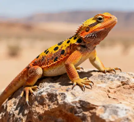
The swelling of the cavalry and other cavities tends to worry and puzzle the owner: is it a joint problem caused by a gout or an accidental fracture? There are significant differences in treatment strategies, care priorities and prognosis. This paper presents an integrated programme that can be implemented and that is as “innocuous as possible”, ranging from common causes, identifying elements, to specific response steps. It should be emphasized that such cases fall within the realm of clinical judgement and that it is advisable to bring a pet to a veterinary service with a reptile specialty as soon as possible.
The content focuses on common causes and directions: aphrodisiac arthritis (heavy swollen arthritis due to hyperureasis), fractures or distortions, localized infections or sepsis, soft tissue contusion, arthritis, etc. The focus and risk of care for each cause are different. At the heart of the diagnostic path: medical history and behaviour, body examin, image (X-line, CT if necessary) and seros/seroure routines. It must not be judged solely by appearance. Response principles and measures: to stabilize the environment and moisture, to observe records, to request veterinary doctors to develop a medical programme; to prohibit the use of self-pharmaceuticals and, in particular, to refrain from injecting drugs (e.g. painkillers/inflammants) in order to avoid aggravating conditions. Long-term management: reasonable nutritional ratio to calcium phosphorus, adequate moisture, moderate activity, periodic review to avoid repeated exposure to chronic joint damage.
The symptoms of the phenomenon identification point (the focus of household observations) are often characterized by lower symmetric swelling in the vicinity of one or more major joints, limited but not immediately deformed activity. The skin temperature in the swelling area may be slightly higher and the pain is associated, but in most cases it is not accompanied by visible openings. There may be dietary transport history: chronic high protein, uneven calcium phosphorus, inadequate drinking water, etc. Recent increases in body weight and abdominal stress are also of concern if they are associated with the kidney function burden. Visible malformations, non-weighting of limbs, rapid swelling and intense pain during touching after fracture/misplacement. It is accompanied by movement restrictions and unusual positions, with the possibility of early signs of muscle atrophy. Other common causes include local infections, sepsis, soft tissue contusions, etc., which are often accompanied by local heat pains, secretions and skin fractures. Arthritis, rampitis, etc. can also cause swelling but often chronic trends.
Phased, implementable first phase of a complete response: the family ' s steadyness and first aid chain (within 24-48 hours) maintains a moist and warm base area to avoid sudden temperature changes. The suitable daytime temperature zone for the palace should be as stable as possible to avoid overcooling or overheating. Provide clean water and fresh diets to ensure that water can be replenished at any time, with short periods of shallow tub bathing if necessary (temperature water, suitable to cover body joints, short periods to avoid stress). Restraint and stress sources to reduce movement and severe shaking. In the early stages of the swelling, the body was kept as quiet as possible, avoiding self-attacking or stepping on the injured part. Key messages are recorded: swelling, changes in size, relative symmetry, apparent restrictions on activity associated with it, recent diets and drinking water, history of trauma, exposure to excessive protein feed, etc.
Phase II: A veterinarian with reptile experience to visit as soon as possible and to complete a preliminary diagnostic appointment, giving priority to the screening of X-rays and the necessary blood tests. At the time of the visit: time lines for the onset of the disease, recent food and water intake, trauma, other companion pets, pain signals you observe (e.g. refusal to move, quick flight, scratching, etc.). Diagnosis processes may include: medical examination, X-line images (bone fracture/fault, joint cavity or fracture trail), blood tests (calcium, blood urea, liver renal function, white cell count, etc.), joint fluid piercing tests (e.g., treatment of infectious inflammation). When the diagnosis is clear, treatment is carried out under the veterinary programme. Professional advice is required for pain management, inflammation control, infection control and nutrition adjustment.
Phase III: Treatment and care (based on veterinary prescriptions) aphrodisiac arthritis: usually focused on correcting metabolic abnormalities, improving hydrosis, adjusting feed ratios. Veterinary doctors may recommend adjusting the sources of protein, increasing the balance of alkaline minerals and reducing inflammation and pain with appropriate drug programmes. Any drug should be used under veterinary guidance. Fractures/faults: Depending on the type of fracture, treatment varies from conservative fixation to intervention treatment (e.g. external fixation, stitching or surgery) if necessary. It is forbidden to fix or bind itself in order not to oppress or affect blood circulation. Infective Inflammation: Antibiotics and local care, clean-up and, if necessary, septic and wound care. Elements of long-term care: calcium phosphorus ratio (e.g., common bone problems associated with calcium intake, adjustment of calcium dosage to vitamin D3 as recommended by veterinary doctors), water management, balance of daily activity, avoidance of metabolic disorders caused by chronic high salt or fat diet.
Phase IV: Long-term management of diet and nutrition in daily life: provision of a balanced diet for ceremonial health and observance of absolute taboos (e.g., risks of pathogens in the raw meat, sources of pollution). Ensure that calcium is moderate and, if necessary, add composite calcium/vitamin D3 formulations to veterinary instructions. To avoid the long-term feeding of high proteins and indigestionable foods. Humidity and humidity: water sources are clean and periodically replaced; proper increase in humidity contributes to joints and skin health, but to avoid environmental humidity leading to caricature and skin infections. Environmental management: Provide controlled temperature gradients, hideouts, climbing structures, reduce stress and ensure proper weight distribution of limbs in daily activities. Regular review: Reconfirmation of post-operative/treatment plans, review of image and blood test indicators, monitoring of changes in indicators such as urine acid, calcium, kidney function and prevention of chronic complications. Observation records: Daily observation logs are established to record feeding, drinking water, activity, swelling changes, defecation etc., so that doctors can assess the effects of treatment.
The risks requiring attention and the risk of inhibition of self-use are extremely high, especially when people use painkillers or non-paralytic anti-inflammatory drugs (NSAIDs) for creeping can cause serious gastrointestinal haemorrhage, kidney damage and even fatality. Any drugs should be prescribed by veterinary medicine. There should be no blind holding, oppression or towing of limbs without visual evidence. Medical treatment is required as soon as possible in cases of acute respiratory activity, abnormal pulses, continued food denial for more than a few days, apparent loss of body weight, excretion abnormalities, etc.