The case is that of a 12-year-old male sterilised domestic cat with 2 weeks of respiratory difficulties and a few days of an appetite. Cat breast anomalies may be difficult to explain. Can you find an anomaly and list some identified diagnosis?
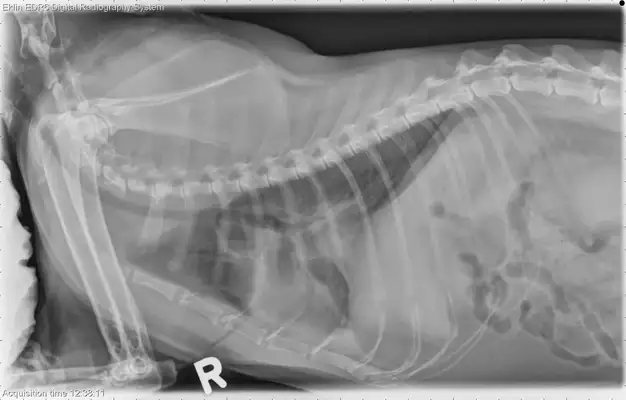
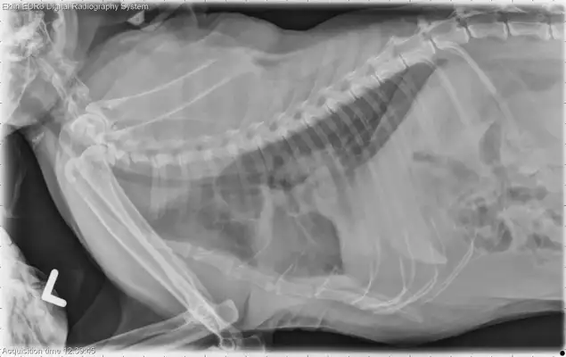 Imaging the results.
The shape of the heart shadow is normal, the lungs are normal. The sub-pulmonary leaves or transected areas have increased the opacity of soft tissue, overlapped with the aerobic contours and covered the back of the heart. Fragments of the soft tissue can be seen between multiple lung leaves. There is a slight bronchial pattern throughout the lungs. The musculoskeletal structure is not significantly abnormal. Identification diagnosis
Cervical cavity (emission fluid, leaching fluid, blood, emulsions) with by-pulmonary leaves or tails (tumours, bulges, septs), possibly subsequent to a swollen bronch pattern of inflammation or representing light water Swollen.
 The full video package of the Veterinary Examination [with downloading] + document material [with small stoves & Zeave video web lessons + mind map + four colour notes + summary of mouths + reality + practice]
The CT image shows an increase around the swelling and contact with both the heart and the muscular muscle. The tail veins are in contact with the swelling, are slightly under pressure, and the edibles move backwards. The diagnosis is still end-of-pipe or pulmonary foe swelling and may be tumours. During the operation it was found that the swelling was attached to the heart pack and the muscular muscles and could not be removed. The post-mortem was diagnosed with sub-pulmonary folic cancer, spreading to muscular and pericardium.
Original by AllISON ZWINGENBERGER